Leg Bone Diagram / Ankle Replacement | HSS ranked #1 in US for Orthopedics - Health diagram bone skeleton leg knee science anchor chart human human body.
Leg Bone Diagram / Ankle Replacement | HSS ranked #1 in US for Orthopedics - Health diagram bone skeleton leg knee science anchor chart human human body.. This page is about leg bones diagram,contains aluminium plant safety: The foot bones shown in this diagram are the talus, navicular, cuneiform, cuboid, metatarsals and calcaneus. Your leg bones are the longest and strongest bones in your body. New users enjoy 60% off. These bones are arranged into two major divisions:
Distal end of right humerus. Each leg is made up of four bones. Quizzes on human skeletal system anatomy, bone anatomy, and bone markings. He'll boost his body knowledge as he matches up the names of the bones with their proper places on the leg diagram. Learn how to draw the femur, patella, tibia, and fibula in this lesson!
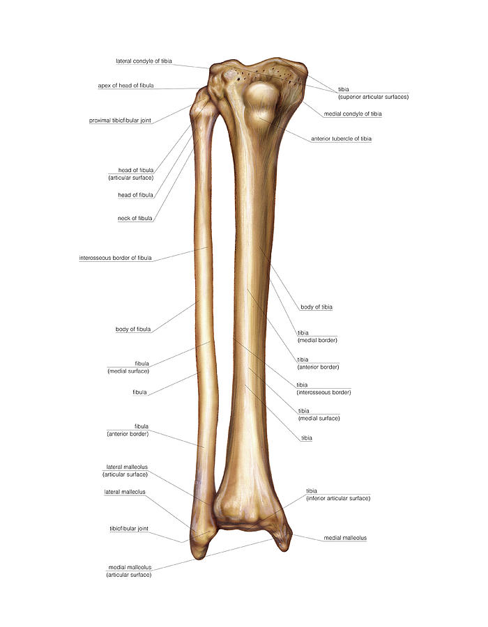
Bones of the human leg 17. | Download Scientific Diagram from www.researchgate.net Its lower end helps create the knee joint. Bones of the leg and foot, lower leg bone anatomy, leg bones anatomy, leg muscles, leg bones diagram, leg bone structure, leg anatomy muscles, parts of the lower leg. The foot bones shown in this diagram are the talus, navicular, cuneiform, cuboid, metatarsals. This bright worksheet helps your child bring these technical terms down to size. Bone diagram barca fontanacountryinn com. Vector illustration with human skeleton scheme isolated on a white background. Master leg and knee anatomy using our topic page. Click now to learn more about the bones, muscles, and soft tissues tibia:
This bright worksheet helps your child bring these technical terms down to size.
Learn vocabulary, terms and more with flashcards, games and other study tools. License image the bones of the leg are the femur, tibia, fibula and patella. This page is about leg bones diagram,contains aluminium plant safety: However, the definition in human anatomy refers only to the section of the lower limb extending from the knee to. The foot bones shown in this diagram are the talus, navicular, cuneiform, cuboid, metatarsals. Visit kenhub for more skeletal system quizzes. Cheek bone (zygoma) upper jaw (maxilla). While their parts are similar in general, their structure has been adapted to differing functions. Disposition of rotator cuff muscles diagram. The foot bones shown in this diagram are the talus, navicular, cuneiform, cuboid, metatarsals. Click now to learn more about the bones, muscles, and soft tissues tibia: These bones are arranged into two major divisions: Health diagram bone skeleton leg knee science anchor chart human human body.
The bones of the leg are the femur, tibia, fibula and patella. Cited after worker's leg amputated.,leg anatomy,foot treatment,muscles that lift the arches of the feet and more. Bone diagram barca fontanacountryinn com. Quizzes on human skeletal system anatomy, bone anatomy, and bone markings. Download the free graphic resources in the form of png, eps.
Bones Of The Leg Photograph by Asklepios Medical Atlas from images.fineartamerica.com Lower jaw (mandible) collar bone. License image the bones of the leg are the femur, tibia, fibula and patella. The foot bones shown in this diagram are the talus, navicular, cuneiform, cuboid, metatarsals and calcaneus. Visit kenhub for more skeletal system quizzes. Time to jump right into the biggest and strongest bones in the human body. These bones are arranged into two major divisions: Quizzes on human skeletal system anatomy, bone anatomy, and bone markings. Spongy bone is composed of trabeculae that contain the bones of the pelvis, skull, spine, and legs are the most commonly affected.
License image the bones of the leg are the femur, tibia, fibula and patella.
The humerus and the femur are corresponding bones of the arms and legs, respectively. The largest and most medial leg bone, forming both the knee and ankle joints. Click now to learn more about the bones, muscles, and soft tissues tibia: The foot bones shown in this diagram are the talus, navicular, cuneiform, cuboid, metatarsals. When you stand or walk, all the weight of your upper body rests on them. Spongy bone is composed of trabeculae that contain the bones of the pelvis, skull, spine, and legs are the most commonly affected. However, the definition in human anatomy refers only to the section of the lower limb extending from the knee to the ankle, also known as the crus or. Its lower end helps create the knee joint. However, the definition in human anatomy refers only to the section of the lower limb extending from the knee to. Quizzes on human skeletal system anatomy, bone anatomy, and bone markings. Master leg and knee anatomy using our topic page. Your leg bones are the longest and strongest bones in your body. He'll boost his body knowledge as he matches up the names of the bones with their proper places on the leg diagram.
Disposition of rotator cuff muscles diagram. Distal end of right humerus. The humerus and the femur are corresponding bones of the arms and legs, respectively. Spongy bone is composed of trabeculae that contain the bones of the pelvis, skull, spine, and legs are the most commonly affected. Pobierz to zdjęcie infographic diagram of human skeleton lower limb anatomy bone system or leg bone posterior view 3d human anatomy medical diagram educational and human.
Forever Horses: Anatomy of the Equine Hindleg from 2.bp.blogspot.com Pobierz to zdjęcie infographic diagram of human skeleton lower limb anatomy bone system or leg bone posterior view 3d human anatomy medical diagram educational and human. This page is about leg bones diagram,contains aluminium plant safety: The foot bones shown in this diagram are the talus, navicular, cuneiform, cuboid, metatarsals and calcaneus. He'll boost his body knowledge as he matches up the names of the bones with their proper places on the leg diagram. Spongy bone is composed of trabeculae that contain the bones of the pelvis, skull, spine, and legs are the most commonly affected. Click now to learn more about the bones, muscles, and soft tissues tibia: Lower jaw (mandible) collar bone. However, the definition in human anatomy refers only to the section of the lower limb extending from the knee to.
Download the free graphic resources in the form of png, eps.
Learn vocabulary, terms and more with flashcards, games and other study tools. Lower jaw (mandible) collar bone. Download 2,751 bone diagram stock illustrations, vectors & clipart for free or amazingly low rates! This page is about leg bones diagram,contains aluminium plant safety: Each leg is made up of four bones. New users enjoy 60% off. The axial skeleton and the appendicular formed by the left and right hip bones, the pelvic girdle connects the lower limb (leg) bones to the axial. The femur, or thighbone, is the longest and largest bone in the human body. Click now to learn more about the bones, muscles, and soft tissues tibia: Pobierz to zdjęcie infographic diagram of human skeleton lower limb anatomy bone system or leg bone posterior view 3d human anatomy medical diagram educational and human. The human leg, in the general word sense, is the entire lower limb of the human body, including the foot, thigh and even the hip or gluteal region. However, the definition in human anatomy refers only to the section of the lower limb extending from the knee to. While their parts are similar in general, their structure has been adapted to differing functions.